When the physical wave comes upon a border between two various resources (different traditional impedance) power is reflected back again to the ultrasound transducer.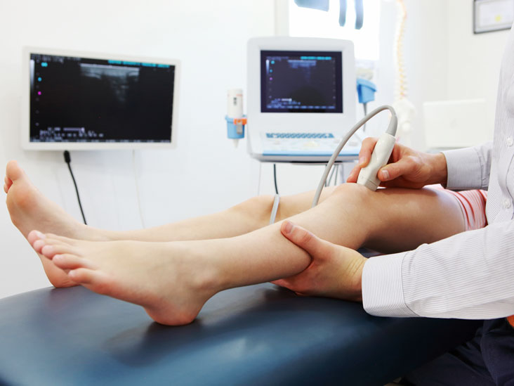
That reflected power reveal is found by theprobe. Enough time it takes for the reveal to visit back once again to the probe is electronically calculated and applied to estimate, as well as to display, the level of the tissue. The frequencies useful for medical ultrasonography are usually in the product range of 1 to 15 MHz. Larger wavelengths have a lower wavelength, providing images with a larger resolution. Nevertheless, the reduced total of the wave is improved at higher frequencies, so to be able to most efficiently enter greater tissues, a lowered frequency (3-5MHz) is used.
Ultrasound transducers make images of muscle and soft tissue, helpful for defining borders between stable and fluid-filled spaces. Ultrasound transducers give live photographs, allowing operators to pick the most readily useful parts for rapid diagnoses. They show the design and purpose of central organs. It offer a good method to examine the musculoskeletal process to identify problems with muscles, ligaments, tendons and joints. In addition it guide in distinguishing obstructions, stenosis and different vascular abnormalities.
Modern, high-class ultrasoundsystems use the most readily useful ultrasound in tarneit transducers technology, coupled with outstanding processors and an easy to use interface. The picture quality depends primarily on the ultrasound transducer, that is leading conclusion that transmits and receives the technical energy. Modern ultrasound transducers give users unparalled and unparalleled multi-modality ultrasound experience.
Carotid ultrasonography, applied to evaluate body flow to the carotid arteries, in addition to the intra-cerebral arteries. Echocardiography, which can be an ultrasound that shows the motion of one’s heart, while the muscle dilates and contracts. Disaster medical experts frequently make use of a kind of probe. Also, it’s found in the ER as a routine method of rapidly assessing the cause of a patient’s abdominal pain.
Urologists often use ultrasound transducers to detect the amount of water that is in a patient’s bladder. Gynecologists might perform pelvic sonogram utilizing an ultrasound to see a picture of the pelvic floor in women and spot any abnormalities. More in depth photographs of the tendons, nerves, muscles, ligaments and different smooth muscle places that might have been affected by damage or trauma. Arterial probe is used by cardiologists to check for possible obstructions in the arteries, or even to detect DVT. In gastroenterology, health practitioners can see the abdomen using an probe to see organs like the aorta, pancreas, gall kidney, kidneys, liver and spleen.
There are four types of accessible ultrasound image. The option which kind of image to make use of is dependent upon the goals for a specific check, the phenomena being investigated and what gear is available. The most common and form of ultrasound picture is a series of smooth, two-dimensional corner part photos of the scanned tissue. Described simply as 2d ultrasound, this setting of reading is still standard for several diagnostic and obstetric scenarios after a half-century of use.
In recent years, 2d photographs have also been projected into three-dimensional representations. This really is achieved by scanning structure cross areas at numerous sides and reconstructing the info obtained into a three-dimensional image. A standard use for 3d ultrasound pictures is to provide a more complete and realistic image of a developing fetus. By upgrading 3d ultrasound photos in rapid sequence, sonographers may also create 4d ultrasound pictures. In the 4d ultrasound, the last aspect, time, adds movement and generates the most sensible illustration of all.
Sometimes, 3d and 4d ultrasound photographs might show abnormalities not commonly seen using second ultrasound. For expectant mothers and members of the family, the capability to see reasonable photographs of an unborn child in the womb could be rewarding and heartwarming although the medical neighborhood generally warns against performing ultrasound tests solely with this purpose.